Radiation Heatmap & Tumor Detection
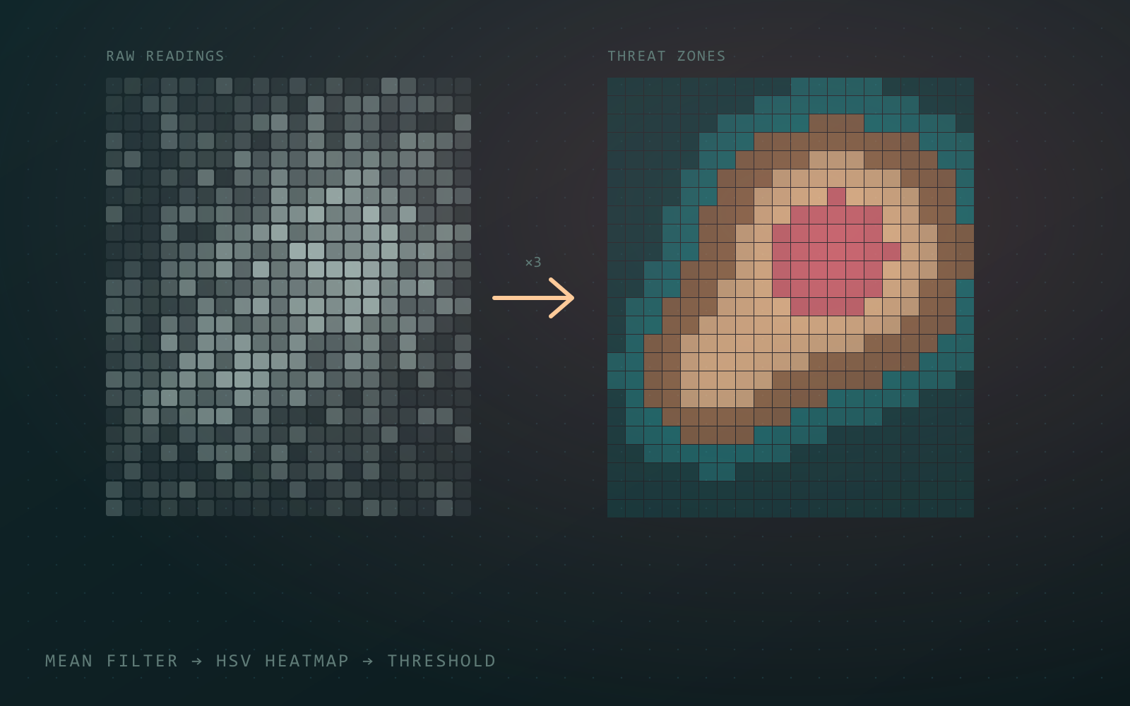
One image-processing pipeline, two uses. Noisy radiation readings are smoothed with a mean filter and mapped onto terrain imagery as colour, and the same filtering flags bright regions in brain scans as possible tumors.
- Stack
- MATLABImage ProcessingHSV ColourUnit Testing
- Highlights
- Three-pass n×n mean filter for noise removal
- Smooth heatmap and discrete threat zones via the HSV hue channel
- Threshold-based tumor flagging on filtered scans
The problem
Raw sensor readings are noisy, and a grid of numbers means little to the person reading it. The job is to clean the data, then show it in a way people can take in at a glance.
Approach
Denoise. An n×n mean filter is applied three times with replicated edges, which smooths out spikes without shrinking the image.
Heatmap and zones. The image is converted to HSV and the hue channel is set from the radiation value: a smooth gradient for the heatmap, and fixed hue bands for discrete threat zones. Saturation is maxed so the colour reads over the terrain.
Tumor detection. The same filter is run over a brain scan, and any region still brighter than a tuned threshold is flagged for follow-up. The threshold was chosen by testing values against sample scans.